
Learning Objectives
- [AHL] Explain the directionality of RNA and DNA.
- [AHL] Describe the role of purine-to-pyrimidine bonding in maintaining DNA helix stability.
- [AHL] Identify the structure and function of a nucleosome.
- [AHL] Analyse the evidence from the Hershey-Chase experiment supporting DNA as the genetic material.
- [AHL] Interpret Chargaff’s data on the relative amounts of pyrimidine and purine bases across diverse life forms.
Part 1: Directionality of RNA and DNA
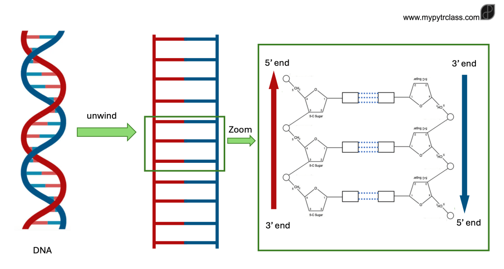
Directionality of RNA and DNA in Enzymatic Processes
- Directionality affects key biological processes, ensuring correct molecular interactions:
- Replication – DNA polymerases copy DNA.
- Transcription – RNA polymerase creates an RNA copy of DNA.
- Translation – Ribosomes read RNA to determine the amino acid sequence of a polypeptide.
- Correct orientation is essential for DNA and RNA strands to fit into the active sites of enzymes and ribozymes.
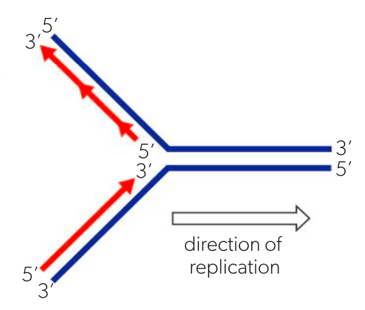
Replication
- DNA nucleotides are added to the 3′ end of the growing strand.
- The 5′ phosphate of a free nucleotide binds to the 3′ deoxyribose sugar of the existing strand.
- DNA replication occurs in the 5′ to 3′ direction.
- Both DNA strands serve as templates, but since they are antiparallel:
- One strand is synthesized in the same direction as replication.
- The other strand is synthesised in the opposite direction, leading to differences in the process.
Transcription
- RNA nucleotides are added to the 3′ end of the growing RNA strand.
- The 5′ phosphate of a free nucleotide links to the 3′ ribose sugar of the existing strand.
- Transcription also occurs in the 5′ to 3′ direction.
- Only one of the two DNA strands is used as a template for RNA synthesis.
- The RNA strand is assembled in the same direction as transcription proceeds.
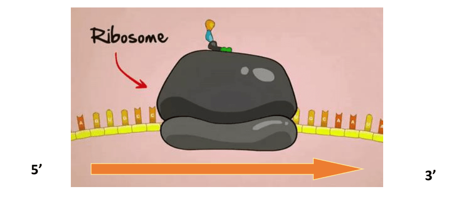
Translation
- RNA carries the genetic instructions for building a polypeptide.
- The ribosome moves along the RNA strand from 5′ to 3′, linking amino acids in sequence.
- Translation follows the 5′ to 3′ direction just like replication and transcription.
Part 2: Maintaining DNA Helical Structure
Purine-pyrimidine
- Nitrogenous bases in DNA are classified into two groups:
- Purines
- Adenine (A) and Guanine (G)
- Have two rings of atoms.
- Pyrimidines
- Cytosine (C) and Thymine (T)
- Have one ring of atoms.
- Purines
- Base pairing in DNA follows a strict purine-to-pyrimidine rule:
- For each base pair, if one is a purine and one must be complimented with a pyrimidine.
- This ensures equal width for all base pairs, maintaining a uniform structure in the double helix.
- The consistent spacing stabilises the DNA molecule and allows for any base sequence in genes.

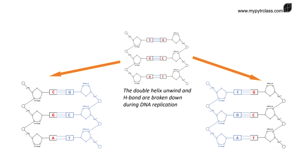
Chargaff’s rules
- Base Pairing Rule
- Amount of A = amount of T and,
- Amount of C = amount of G
- This provided evidence for complementary base pairing in DNA.
- Purine-Pyrimidine Balance
- A and G are purines, and C and T are pyrimidines
- Chargaff’s data showed that:
- Total purine content = total pyrimidine content in a DNA sample.
- Species Variation
- A = T and C = G ratios remained constant within a species
- The proportions of bases varied between species.
- This suggested that DNA composition is unique to each organism, supporting its role as the genetic material.
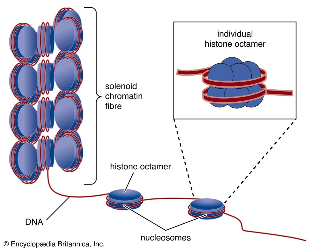
Part 3: What are Nucleosomes?
DNA is packed (condensed) in the nucleus in a form of chromosomes. To do this, the DNA must wrap around many proteins by coiling them. For each 2 coil, the DNA strand wraps 2 copies 4 different histone proteins. These proteins are called octamer. The beginning and the end of the coil will then be “held tight” by another protein called the H1 protein.

Nucleosome
A DNA stand wrapping 2 copies of 4 different histone proteins (octamer) and locked or reinforced by H1 protein
- Eukaryotes (plants, animals, and other eukaryotic organisms) have nucleosomes, where DNA is wrapped around histone proteins.
- Bacteria lack nuclei and histones, meaning their DNA is “naked” and not associated with proteins for packaging.
Chromosomes as Condensed DNA
- Chromosomes are highly condensed forms of DNA, allowing genetic material to fit within the cell nucleus.
- DNA condenses by wrapping around histone proteins, forming nucleosomes, which further coil and fold into chromatin.
- During cell division (mitosis and meiosis), chromatin condenses even further to form distinct chromosomes, making genetic material easier to separate.
- Chromosome structure ensures accurate DNA replication and distribution to daughter cells.
Steps in DNA Condensation
- Nucleosomes formation
- DNA stand wraps histone octamer and reinforced by H1 protein
- Solenoid formation
- The nucleosomes coil to form a loop or a spring structure
- Chromatin fibre formation
- Solenoid fibre further condense by coiling and looping about a scaffolding protein
- Two types of chromatin:
- Euchromatin: Loosely packed – available for gene expression i.e transcriptionally active.
- Heterochromatin: Densely packed – unavailable for gene expression i.e transcriptionally inactive.
Part 4: Evidence for DNA as Genetic Material
Hershey–Chase Experiment: Evidence for DNA as Genetic Material
- Scientists knew chromosomes played a role in heredity but were unsure if DNA or protein was the genetic material.
- Protein was initially considered the better candidate due to its complexity (20 amino acids vs. 4 nucleotide types in DNA).
- Experiment Setup:
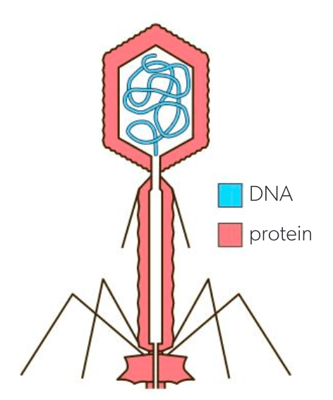
- Organism Used: T2 bacteriophage (a virus with a protein coat and DNA inside).
- Key Idea: Viruses inject genetic material into host cells to replicate.
- Radioactive Labeling:
- 35S (Sulfur) labeled protein (proteins contain sulfur but not phosphorus).
- 32P (Phosphorus) labeled DNA (DNA contains phosphorus but not sulfur).
- Method:
- Viruses with labeled protein or DNA infected bacteria.
- A blender separated viral coats from bacteria.
- Centrifugation concentrated bacteria into a pellet.
- Radioactivity was measured in the pellet (bacteria) and supernatant (viral coat).
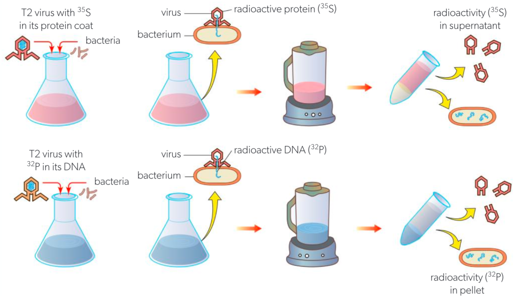
Process of the Hershey–Chase experiment
- Results & Conclusion:
- Radioactive phosphorus (32P) was found inside the bacteria, proving DNA entered the cells.
- Radioactive sulfur (35S) remained in the viral coat, meaning protein did not enter the cells.
- Conclusion: DNA, not protein, is the genetic material responsible for viral replication.
Questions
- Differentiate between a supernatant and a pellet. [2]
- Justify why the genetic material should be located in the pellet rather than the supernatant. [2]
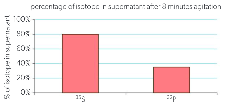
- Identify the percentage of ³²P that remains in the supernatant. [1]
- Calculate the percentage of ³²P that is found in the pellet after centrifugation. [2]
- Evaluate the evidence supporting DNA as the molecule responsible for transforming bacteria into infected cells. [3]
![ESS 8.1.3 [AHL] Biocapacity and Environmental Migration](https://mypytrclass.com/wp-content/uploads/2025/12/image-4.png?w=1024)


![ESS 8.3.4 [AHL] Photochemical Smogs and Tropospheric Ozone](https://mypytrclass.com/wp-content/uploads/2025/12/image-1.png?w=1024)