
Learning Objectives
- Explain the chemical diversity of R-groups in amino acids and how they contribute to the vast diversity in protein structure and function
- Describe the impact of primary structure on protein conformation, emphasising how the sequence of amino acids determines protein shape and function
- Identify the pleating and coiling patterns that form the secondary structure of proteins, including alpha-helices and beta-pleated sheets
- Explain how the tertiary structure of proteins depends on hydrogen bonds, ionic bonds, disulfide covalent bonds, and hydrophobic interactions
- Analyse the effect of polar and non-polar amino acids on protein folding and stability in tertiary structures
- Differentiate between the quaternary structure of non-conjugated and conjugated proteins, and explain their structural significance
- Compare the form and function of globular and fibrous proteins, highlighting how their structures relate to their biological roles
Part 1: The R Group of Amino Acids
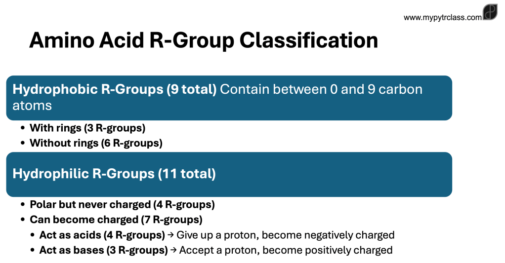
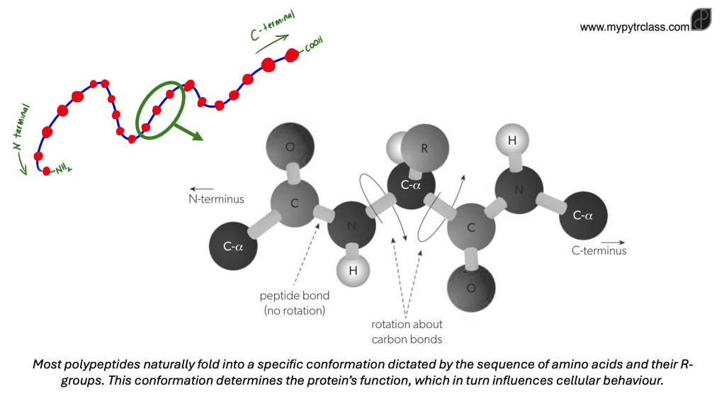
The 20 amino acids used by ribosomes to form polypeptides vary significantly in the chemical nature of their R-groups. When amino acids link into a polypeptide, their amine and carboxyl groups form peptide bonds, leaving a free amine group (–NH₂) at one end and a carboxyl group (–COOH) at the other.
- The hydrogen atom attached to the alpha carbon has minimal impact on the polypeptide’s properties
- The R-groups determine chemical characteristics of the polypeptide.
Some R-groups are hydrophobic, while others are hydrophilic, with some being polar and others carrying a charge (+ or −) due to their acidic or basic nature. This diversity in R-groups enables organisms to produce a vast range of proteins with specialized functions.
| Elements in R-group | Number of Amino Acids |
|---|---|
| H only | 1 |
| C and H only | 5 |
| C, H, and S only | 2 |
| C, H, and N only | 5 |
| C, H, and O only | 5 |
| C, H, N, and O | 2 |
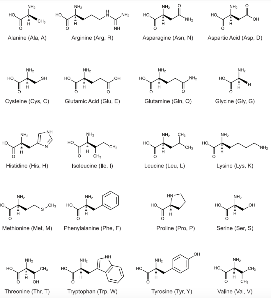
In some proteins, amino acids extend beyond the standard 20, typically due to modifications after polypeptide synthesis. One example is collagen, a structural protein that provides tensile strength to tendons, ligaments, skin, and blood vessel walls. Ribosomes initially incorporate proline at multiple positions within collagen polypeptides, but some of these prolines are later converted into hydroxyproline, enhancing collagen’s stability.
Part 2: Structure of Protein
Primary Structure
- A chain of polypeptide with no shape. The tetrahedral domain of the alpha C atoms allow rotation of the sigma bonds (single bond) about the alpha C
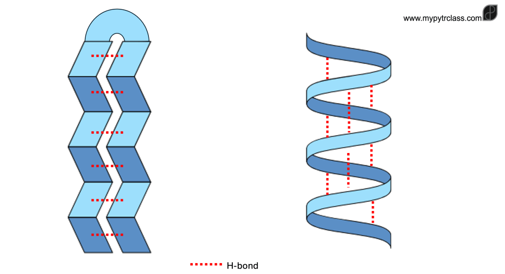
Secondary Structure
- The C=O and N-H can interact and form H-bonding between two amino acids.
- As the result, two general 2D shapes are obtained:
- α-helix
- The polypeptide is coiled into a helical shape, with hydrogen bonds forming between adjacent turns of the helix.
- β-pleated sheet
- Two or more segments of the (one) polypeptide align parallel to each other, with hydrogen bonds connecting them. These segments run in opposite directions, creating a pleated appearance due to tetrahedral bond angles.
- α-helix
Tertiary Structure
- The R groups of each amino acid along a polypeptide chain may interact with each other and form different types of stabilising interactions. These include:
- Ionic Bonds
- Formed between positively and negatively charged R-groups.
- Amine groups gain a proton (+H⁺) to become –NH₃⁺, while carboxyl groups lose a proton to become –COO⁻.
- Since these bonds involve protons, they are highly sensitive to pH changes.
- Hydrogen Bonds
- Occur between polar R-groups.
- A hydrogen atom, covalently bonded to an electronegative atom like oxygen (O) or nitrogen (N), forms a weak attraction with another electronegative atom, stabilising the protein’s shape.
- Disulfide Bonds
- Strong covalent bonds that form between two cysteine amino acids, making them the most stable type of interaction in protein folding.
- Hydrophobic Interactions
- Non-polar R-groups cluster together away from water, helping to stabilise the protein’s structure.
- Ionic Bonds
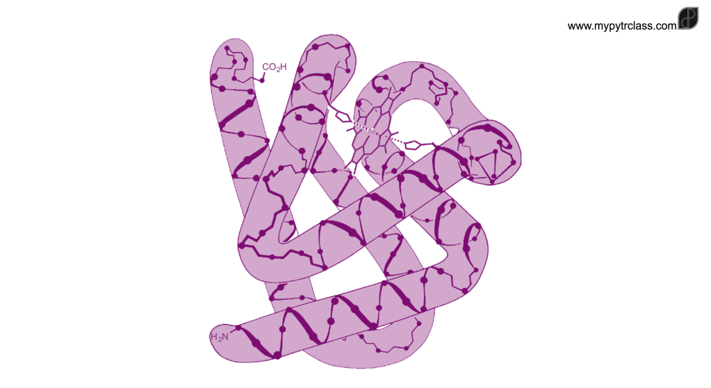
As a polypeptide is synthesised by the ribosome, it begins to fold into its tertiary structure. In some cases, chaperone proteins assist in this process to ensure the protein attains the correct conformation and becomes fully functional. A diverse range of 3D shapes is produced, with most proteins adopting a globular structure. Within these folded proteins, regions of secondary structure such as α-helices and β-pleated sheets are often present.
However, some polypeptides do not fold into a compact shape and instead remain elongated, lacking a distinct tertiary structure. These proteins, known as fibrous proteins, play structural roles in the body.
Quaternary Structure
Quaternary structure of non-conjugated and conjugated proteins
| Type of Protein | Composition | Quaternary Structure | Examples | Function of Non-Polypeptide Components |
|---|---|---|---|---|
| Non-Conjugated Proteins | Only polypeptide subunits | Formed by interactions between polypeptides (e.g., disulfide bonds, hydrogen bonds) | – Insulin (two polypeptides linked by disulfide bonds) – Collagen (three polypeptides wound together) | – No non-polypeptide components |
| Conjugated Proteins | Polypeptides + one or more non-polypeptide subunits | Includes non-polypeptide subunits in addition to polypeptides | – Hemoglobin (four polypeptides + heme groups) – Many enzymes with non-polypeptide components | – Non-polypeptide components (e.g., heme group in haemoglobin) contribute to functionality, such as oxygen binding or catalytic activity. |
Part 3: Globular and Fibrous Protein
| Type of Protein | Structure | Function | Examples |
|---|---|---|---|
| Fibrous Proteins | – Elongated polypeptides without typical tertiary folding – No secondary structures like alpha helices – Quaternary structure formed by polypeptide chains linked into fibers with hydrogen bonds | – Provide structural support and strength – Form fibers or filaments for tissues requiring high tensile strength | – Collagen: Triple helix structure formed by three polypeptides – Functions in skin, tendons, ligaments, cartilage, and the eye |
| Globular Proteins | – Rounded shape formed by folding polypeptides – Intricate 3D shape stabilized by R-group interactions | – Function based on precise conformation for specific interactions – Enzyme active sites and receptor binding sites | – Enzymes: Specific shape allows catalysis of biochemical reactions – Insulin: Binds to specific receptors to regulate blood sugar levels |
![ESS 8.1.3 [AHL] Biocapacity and Environmental Migration](https://mypytrclass.com/wp-content/uploads/2025/12/image-4.png?w=1024)


![ESS 8.3.4 [AHL] Photochemical Smogs and Tropospheric Ozone](https://mypytrclass.com/wp-content/uploads/2025/12/image-1.png?w=1024)